Illustrative Descriptions of Organelles

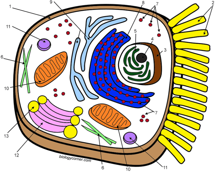
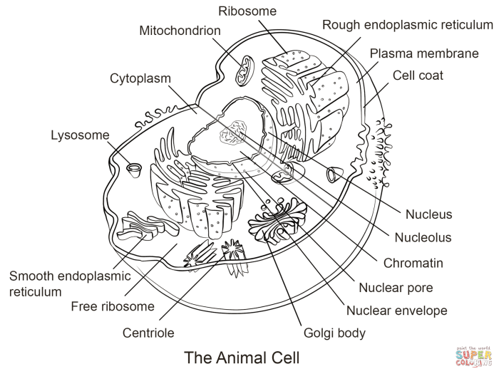
Biology corner animal cell coloring – The following descriptions aim to paint a vivid picture of the animal cell’s internal landscape, focusing on the visual characteristics and functions of its major organelles. Imagine each organelle as a meticulously crafted component within a bustling, miniature city, each playing a crucial role in maintaining the overall health and function of the cellular metropolis. Accurate representation of these organelles in your coloring exercise will require careful attention to their size, shape, and internal structures.
Cell Nucleus
The nucleus, the cell’s control center, is typically the most prominent organelle, a large, roughly spherical structure occupying a significant portion of the cell’s volume. Picture it as a dark, dense sphere, often centrally located, but sometimes pushed to the side by other organelles. Its defining feature is the nuclear envelope, a double membrane punctuated by nuclear pores—tiny gateways regulating the passage of molecules in and out.
Within this envelope lies the chromatin, a diffuse network of DNA and proteins, which condenses into visible chromosomes during cell division. For coloring, depict the nucleus as a darker shade than the surrounding cytoplasm, and subtly indicate the nuclear envelope’s double membrane structure.
Ribosomes
These tiny, granular structures are the protein factories of the cell. While individually too small to see clearly without powerful magnification, ribosomes are often clustered together in large numbers, giving them a somewhat speckled appearance under a microscope. They appear as small dots, either freely scattered in the cytoplasm or attached to the endoplasmic reticulum. When coloring, represent them as numerous, small, dark dots, scattered throughout the cytoplasm and clustered along the rough endoplasmic reticulum.
Their small size and numerous presence should be reflected in the coloring.
So, you’re all about biology corner animal cell coloring? That’s pretty hardcore, I’ll give you that. But sometimes, even a cell needs a break from the nucleus! Need a change of pace? Check out these awesome coloring pages realistic animals for a dose of furry, feathery, or scaly fun before diving back into the intricate world of organelles.
Then, once you’ve had your fill of realistic lions and tigers, it’s back to those amazing animal cell diagrams!
Endoplasmic Reticulum
The endoplasmic reticulum (ER) is an extensive network of interconnected membranes, forming a labyrinthine system throughout the cytoplasm. Imagine it as a vast, folded sheet, extending throughout the cell. There are two types: rough ER and smooth ER. Rough ER, studded with ribosomes, appears as a network of interconnected, slightly darker membranes with a rough texture due to the attached ribosomes.
Smooth ER, lacking ribosomes, presents a smoother, less textured appearance. In your coloring, differentiate the rough and smooth ER by showing a difference in texture; the rough ER should appear more granular than the smooth ER.
Golgi Apparatus
The Golgi apparatus, or Golgi body, resembles a stack of flattened, membrane-bound sacs, or cisternae. Think of it as a series of interconnected pancakes, each slightly curved. These sacs are involved in processing and packaging proteins and lipids. When coloring, depict it as a stack of slightly curved, flattened sacs, with a lighter color than the nucleus.
The layering and the slightly curved nature of each sac should be carefully rendered.
Mitochondria
Mitochondria, the powerhouses of the cell, are sausage-shaped or oval organelles with a double membrane structure. They are relatively large, easily visible under a microscope. The inner membrane is folded into cristae, creating a significantly increased surface area for energy production. When coloring, represent them as oval or sausage-shaped structures with a darker inner membrane folded into cristae, to highlight the increased surface area.
Their relatively large size should be clearly shown in relation to other organelles.
Lysosomes
Lysosomes are small, membrane-bound sacs containing digestive enzymes. They appear as small, spherical vesicles, scattered throughout the cytoplasm. For coloring, represent them as small, dark, round structures, reflecting their role in waste disposal and cellular recycling. Their small size compared to the nucleus and mitochondria should be accurately depicted.
Vacuoles
Vacuoles are membrane-bound sacs that store various substances. In animal cells, they are generally smaller and more numerous than in plant cells. They appear as clear, membrane-bound sacs of varying sizes and shapes, depending on their contents. In your coloring, depict them as clear or lightly colored sacs, potentially showing some variation in size and shape to reflect their diversity in contents.
Expanding the Activity
The simple act of coloring an animal cell diagram can be a gateway to a deeper understanding of cellular biology. Moving beyond the basic coloring exercise opens up a world of engaging activities that solidify learning and foster critical thinking. By incorporating additional elements, we can transform a passive activity into an active, enriching learning experience. The key lies in creating opportunities for exploration and application of the knowledge gained through visual representation.The coloring worksheet serves as a foundation, a visual map of the cell’s internal landscape.
Building upon this foundation, we can introduce activities that challenge students to think critically about the functions of each organelle and their interrelationships. This transition from passive observation to active engagement is crucial for long-term retention and a deeper comprehension of the subject matter. This approach fosters a more dynamic and meaningful learning process, going beyond simple memorization.
Supplemental Activities
A range of supplemental activities can significantly enhance the learning experience derived from the coloring worksheet. These activities can cater to diverse learning styles, encouraging active participation and knowledge consolidation. Consider these possibilities:
- Labeling Exercise: Students can label the organelles on their completed coloring sheets, reinforcing their understanding of each organelle’s name and location.
- Function Matching: Prepare a list of organelles and their functions, asking students to match each organelle to its correct function. This exercise tests comprehension of the roles each organelle plays within the cell.
- Comparative Analysis: Introduce a plant cell diagram for comparison. Students can identify similarities and differences between animal and plant cells, highlighting unique organelles and structures.
- Cell Analogy Project: Students can create analogies to explain the function of each organelle. For example, the cell membrane could be compared to a city’s border control, while the mitochondria could be compared to power plants.
- Create a Cell Model: Students can construct a three-dimensional model of an animal cell using readily available materials, further solidifying their understanding of the cell’s structure and the spatial relationships between organelles.
Short Quiz
A concise quiz can effectively assess students’ understanding of animal cell structures and functions following the coloring activity. The quiz should focus on key concepts and avoid overly complex questions. Here are some example questions:
- Identify the organelle responsible for energy production. (Answer: Mitochondria)
- What is the function of the cell membrane? (Answer: To regulate the passage of substances into and out of the cell.)
- Which organelle contains the cell’s genetic material? (Answer: Nucleus)
- Describe the role of the ribosomes in protein synthesis. (Answer: Ribosomes are the sites of protein synthesis, translating genetic information from mRNA into proteins.)
- What is the function of the Golgi apparatus? (Answer: The Golgi apparatus modifies, sorts, and packages proteins and lipids for secretion or delivery to other organelles.)
Supplemental Resources
Providing access to additional learning resources expands the learning opportunity beyond the coloring worksheet and quiz. These resources can cater to diverse learning styles and provide deeper insights into the intricacies of animal cells.
- Interactive Online Simulations: Numerous websites offer interactive simulations that allow students to explore the structure and function of animal cells in a dynamic environment.
- Educational Videos: Videos provide a visual and auditory learning experience, explaining complex concepts in an engaging manner.
- Reference Books and Textbooks: These provide detailed information and in-depth explanations of animal cell biology.
- Online Encyclopedias and Databases: Reliable online resources like Wikipedia (used cautiously and critically) or specialized databases offer comprehensive information on specific aspects of animal cell biology.
- Scientific Journals (Age-Appropriate): For older students, access to simplified versions of scientific articles can offer insights into current research and discoveries in cell biology.
Alternative Representations of Animal Cells: Biology Corner Animal Cell Coloring

The humble animal cell, a fundamental building block of life, can be visualized in myriad ways, each offering unique insights and limitations. From the simplicity of a hand-drawn diagram to the immersive experience of a 3D animation, the chosen representation significantly impacts our understanding and appreciation of this microscopic marvel. The selection depends on the intended audience, the level of detail required, and the resources available.Different methods of representing animal cells offer varying degrees of realism and accessibility.
Consider the stark contrast between a meticulously detailed scientific illustration and a child’s playful clay model; both communicate the concept of an animal cell, but with drastically different levels of accuracy and complexity. Choosing the right representation is crucial for effective communication and understanding.
Comparison of Animal Cell Representations
Diagrams, 3D models, and animations each present distinct advantages and disadvantages. Diagrams, often found in textbooks, provide a clear, concise overview of cellular components and their relative positions. However, they lack the three-dimensional context and dynamic aspects of cellular processes. 3D models, conversely, offer a more tangible and spatial understanding, allowing for a more intuitive grasp of the cell’s structure.
Yet, constructing detailed models can be time-consuming and require specialized materials. Animations, while highly engaging and visually stimulating, can sometimes oversimplify complex cellular mechanisms or become overwhelming with excessive detail. The optimal method is always context-dependent.
Advantages and Disadvantages of Different Representations
A simple diagram, for instance, excels in its clarity and ease of reproduction. Its simplicity makes it ideal for quick learning and basic understanding. However, this simplicity also limits its ability to convey the intricate three-dimensional relationships within the cell. The lack of depth can lead to a flattened, inaccurate perception of cellular architecture. In contrast, a meticulously crafted 3D model allows for a much more immersive experience, fostering a deeper understanding of spatial relationships between organelles.
The tactile nature of a 3D model enhances engagement and memory retention. However, creating a highly detailed model can be extremely challenging and may require specialized skills and materials. Finally, animations bring the cell to life, showcasing dynamic processes like endocytosis or protein synthesis. The visual dynamism can greatly enhance understanding, but the complexity of creating accurate animations can be prohibitively expensive and time-consuming.
Creating a Simple 3D Model of an Animal Cell, Biology corner animal cell coloring
Constructing a 3D model provides a hands-on approach to understanding animal cell structure. Using readily available materials, a surprisingly accurate representation can be created. For example, a spherical balloon can serve as the cell membrane. Smaller balloons of varying sizes and colors can represent the nucleus, mitochondria, and other organelles. These can be attached to the larger balloon using glue or tape.
Different colored playdough or modeling clay can also be used to sculpt the organelles, allowing for greater detail and customization. This method allows for a tangible, interactive learning experience, especially beneficial for younger learners or those who learn best through kinesthetic activities. The process fosters creativity and critical thinking as students decide on the size, shape, and placement of each organelle, thereby reinforcing their understanding of cellular structure.
Remember to label each organelle clearly for better understanding and comprehension. This simple model, while lacking the intricate detail of a professional model, effectively conveys the fundamental structure of an animal cell in a visually engaging and memorable way.
