Worksheet Design Considerations

Designing effective and engaging animal cell coloring worksheets requires careful consideration of age appropriateness and learning objectives. The worksheets should be visually appealing and provide opportunities for students to reinforce their understanding of cell structures and functions. We will explore designs suitable for elementary and middle school students, culminating in a comparative worksheet focusing on animal and plant cells.Elementary school worksheets should prioritize simplicity and clarity.
Middle school worksheets can incorporate more detail and complexity, encouraging critical thinking skills. A comparative worksheet will help students understand the key differences between these two fundamental cell types.
Elementary School Animal Cell Coloring Worksheet Design
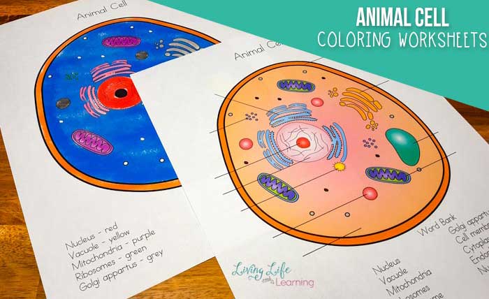
This worksheet focuses on the major organelles of an animal cell, presented in a simplified manner. The design should feature a large, clear Artikel of an animal cell. Each organelle should be clearly labeled with its name, using simple, age-appropriate terminology. For example, instead of “endoplasmic reticulum,” the label might simply read “ER.” The coloring process itself should be straightforward, perhaps assigning different colors to different organelles for easy identification.
The worksheet could also include a small key to help students match colors to organelles. A simple, uncluttered layout will aid understanding and prevent overwhelming the younger learners. The cell should be large enough to allow for easy coloring and labeling within the boundaries of the cell drawing.
Middle School Animal Cell Coloring Worksheet Design
The middle school worksheet builds upon the elementary level by including more organelles and requiring more detailed labeling. This worksheet should present a more complex illustration of an animal cell, perhaps including a cross-section view showing the internal structures in more detail. Organelles should be accurately depicted and labeled with their full names. The worksheet might also include a short answer section requiring students to define the functions of key organelles.
This section could incorporate simple diagrams or flowcharts to help students visualize cellular processes. The overall design should challenge students while remaining engaging and accessible. The addition of a small section on cellular processes could reinforce understanding beyond simple identification.
Comparative Animal and Plant Cell Coloring Worksheet Design
This worksheet allows for a direct comparison of animal and plant cells. It should feature side-by-side illustrations of both cell types, with clear labeling of key organelles. Students should color and label the organelles in both cells, highlighting similarities and differences. To further enhance understanding, a table comparing and contrasting key features of animal and plant cells should be included.
This table should focus on the presence or absence of specific organelles, such as chloroplasts and cell walls, and should use simple language and clear visual cues.
Comparative Table: Animal vs. Plant Cells
| Organelle | Animal Cell | Plant Cell | Key Difference |
|---|---|---|---|
| Cell Wall | Absent | Present (Cellulose) | Provides structural support and protection in plant cells. |
| Chloroplasts | Absent | Present | Site of photosynthesis in plant cells. |
| Large Central Vacuole | Absent or small | Present (Large) | Maintains turgor pressure and stores water and nutrients in plant cells. |
| Shape | Irregular | Usually rectangular or polygonal | Cell wall dictates plant cell shape. |
Visual Aids and Illustrations

Effective visual aids are crucial for understanding complex biological structures and processes. Clear, accurate illustrations enhance comprehension and engagement, particularly in educational materials like coloring worksheets. The following descriptions detail visualizations for key animal cell components and processes.
Animal Cell Illustration
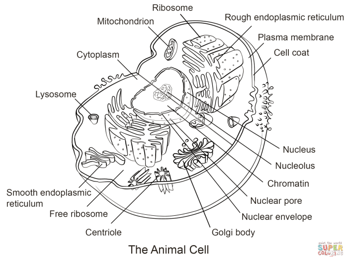
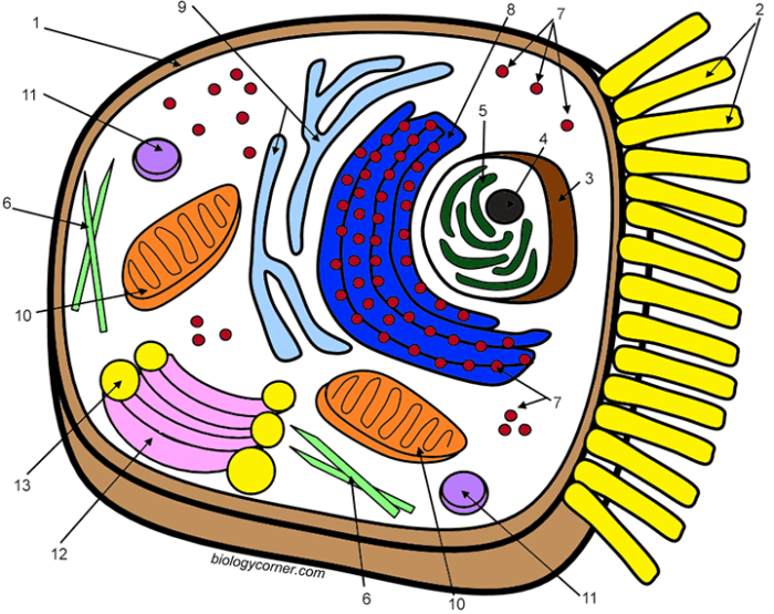
This illustration depicts a typical animal cell, showcasing its various organelles. The cell membrane, a thin, flexible boundary, is represented in a light teal color, subtly highlighting its semi-permeable nature. The cytoplasm, a jelly-like substance filling the cell, is a pale yellow, providing a contrasting background for the organelles. The nucleus, the cell’s control center, is a large, dark purple circle, containing darker purple nucleoli.
The rough endoplasmic reticulum (RER), studded with ribosomes, is depicted as a network of interconnected, light-blue flattened sacs, while the smooth endoplasmic reticulum (SER), involved in lipid synthesis, is shown as a network of pale-green, tubular structures. The Golgi apparatus, responsible for modifying and packaging proteins, is illustrated as a stack of flattened, magenta-colored sacs. Mitochondria, the powerhouses of the cell, are depicted as elongated, crimson-colored structures with internal cristae (folds) indicated by thinner, lighter red lines.
Lysosomes, containing digestive enzymes, are small, dark-orange circles scattered throughout the cytoplasm. Finally, the centrosome, involved in cell division, is represented as a pair of small, dark-green cylinders located near the nucleus. Each organelle is clearly labeled with a concise, legible font in black.
Protein Synthesis Flow Chart
This flow chart visually represents the process of protein synthesis. It begins with a section depicting DNA (deoxyribonucleic acid), shown as a double helix in a deep blue color. An arrow leads to a box labeled “Transcription,” where the DNA sequence is transcribed into messenger RNA (mRNA), represented as a single-stranded molecule in light blue. The mRNA then moves out of the nucleus (indicated by another arrow) and into the cytoplasm.
The next box shows “Translation,” where the mRNA interacts with ribosomes (small, dark-grey spheres), depicted on the rough endoplasmic reticulum (RER). Transfer RNA (tRNA), represented by smaller, light-green cloverleaf shapes, brings specific amino acids to the ribosome. The sequence of amino acids is determined by the mRNA codons. The final box shows the “Polypeptide Chain,” a long chain of amino acids in a light brown color, which folds into a functional protein (represented as a complex, three-dimensional structure in a light-tan color).
Animal cell coloring worksheets are a great way for students to visualize the complex structures within an animal cell. Understanding these structures is key, and checking your work is important; you can find the solutions by looking at the provided answer key for the packet at animal cell coloring packet answers. Then, use the completed worksheet as a reference for future study of animal cell coloring worksheets.
The entire flow chart uses arrows to clearly indicate the direction of the process, ensuring a logical and easy-to-follow sequence.
Cell Membrane Transport
This image illustrates active and passive transport across the cell membrane. The cell membrane is again represented in light teal. Passive transport, which requires no energy, is shown with several small, dark-purple molecules diffusing across the membrane down their concentration gradient, represented by arrows indicating the direction of movement. This section is labeled “Passive Transport (Diffusion).” Active transport, which requires energy, is illustrated with small, dark-green molecules moving against their concentration gradient, with an ATP molecule (adenosine triphosphate), represented as a small, yellow circle, providing the energy for the process.
This section is labeled “Active Transport.” The illustration clearly distinguishes between the two types of transport using different colors and labels, and also depicts the energy requirement for active transport, thus emphasizing the key difference between the two methods.
Worksheet Difficulty Levels: Animal Cell Coloring Worksheets

Creating engaging and educational animal cell coloring worksheets requires careful consideration of difficulty levels to cater to diverse age groups and learning abilities. Differentiation in complexity ensures that all learners, regardless of their prior knowledge or skill level, can benefit from the activity. This involves adjusting the level of detail, the number of structures to identify, and the overall cognitive demands of the task.
Easy Worksheet: Basic Animal Cell Structure
This worksheet focuses on introducing the fundamental components of an animal cell. The design is simple, featuring a large, clearly Artikeld cell with only the most essential organelles: the cell membrane, cytoplasm, and nucleus. The coloring is straightforward, with each organelle assigned a single, easily distinguishable color. For example, the nucleus could be colored purple, the cytoplasm light blue, and the cell membrane dark blue.
This simplicity allows young children to focus on basic identification and color recognition skills. The educational objective is to introduce the basic concepts of cells and their fundamental parts, fostering early familiarity with biological terminology.
Medium Worksheet: Detailed Animal Cell Structure
The medium-difficulty worksheet introduces a more detailed representation of an animal cell. It includes additional organelles such as mitochondria, ribosomes, and the endoplasmic reticulum. The organelles are still clearly delineated, but the level of detail increases, requiring more precise coloring and potentially more nuanced color choices to distinguish between different structures. For instance, the mitochondria could be colored red, the ribosomes small grey dots, the rough endoplasmic reticulum light green, and the smooth endoplasmic reticulum a lighter shade of green.
The educational objective here is to expand upon the basic understanding of cells, introducing more complex structures and their functions, thereby building upon the knowledge gained from the easy worksheet.
Hard Worksheet: Animal Cell Processes and Interactions, Animal cell coloring worksheets
The most challenging worksheet depicts an animal cell actively engaged in various cellular processes. This could include illustrations of processes like protein synthesis, cellular respiration, or endocytosis. The complexity increases significantly, requiring students to not only identify organelles but also understand their roles within the cell’s functional mechanisms. The illustration might depict different organelles interacting with each other, requiring a higher level of cognitive processing and potentially a key to correctly color-code the different stages of a cellular process.
For example, the process of protein synthesis could be illustrated with arrows showing the movement of mRNA from the nucleus to the ribosomes. The educational objective is to encourage deeper understanding of cellular functions and interactions, fostering a more comprehensive grasp of cellular biology.
Adapting Worksheet Design for Different Age Groups
A single worksheet design can be effectively adapted for different age groups and learning abilities by modifying the level of detail and complexity, as described above. For younger children, simpler designs with fewer organelles and easier color schemes are appropriate. Older children or those with more advanced knowledge can benefit from more detailed and complex worksheets that require more cognitive engagement and precise coloring.
The use of accompanying text or labels can also be adjusted based on reading comprehension levels. For example, simpler worksheets could use fewer words or larger font sizes, while more advanced worksheets can incorporate more complex terminology and descriptions.
Answers to Common Questions
What are the benefits of using animal cell coloring worksheets?
They make learning fun, improve knowledge retention through visual aids, and cater to various learning styles.
How can I adapt these worksheets for different learning abilities?
Adjust the level of detail, provide additional support for struggling learners, and offer more challenging extensions for advanced students.
Where can I find printable versions of these worksheets?
The specific availability depends on the source of the worksheets. Many educational websites and resources offer printable versions.
Are there any online resources that complement these worksheets?
Yes, numerous online resources, including interactive simulations and videos, can enhance learning.
