Designing the Coloring Sheet

Animal cell coloring sheet with answers – Creating an engaging and informative animal cell coloring sheet requires careful consideration of both visual appeal and educational accuracy. The design should be clear, concise, and help students easily identify and learn the functions of each organelle. A well-designed sheet will make learning about animal cells fun and memorable.The key to a successful coloring sheet lies in the effective arrangement and clear labeling of the cell’s components.
The visual representation should accurately reflect the relative sizes and positions of the organelles within a typical animal cell, while simultaneously being aesthetically pleasing and easy for children to color.
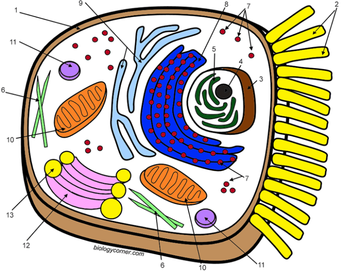
Animal Cell Diagram and Organelle Placement
The animal cell should be depicted as a roughly circular shape, with the nucleus positioned centrally. The nucleus, the largest organelle, should be clearly differentiated from the cytoplasm. Other organelles, such as the mitochondria (depicted as bean-shaped structures), the endoplasmic reticulum (a network of interconnected membranes – represented as a series of interconnected tubes and sacs), the Golgi apparatus (a stack of flattened sacs – represented as layered pancakes), ribosomes (small dots scattered throughout the cytoplasm and on the ER), lysosomes (small, circular organelles), and the vacuoles (smaller and more numerous than in plant cells, shown as small circles), should be strategically placed around the nucleus.
The cell membrane, the outer boundary of the cell, should be a clearly defined line enclosing all the organelles. Each organelle should be clearly labeled with its name, using simple, easy-to-read font.
Coloring Sheet Design and Organelle Differentiation
Each organelle should be assigned a unique color to aid in identification and memorization. For example, the nucleus could be colored purple, the mitochondria blue, the endoplasmic reticulum green, the Golgi apparatus yellow, ribosomes red, lysosomes orange, and vacuoles light brown. The cytoplasm could be a pale yellow or beige. The cell membrane can be a dark Artikel. The coloring sheet should provide ample space around each organelle for coloring without overlapping, ensuring that each organelle is clearly visible and distinguishable.
The labels should be placed near each organelle, but not so close as to interfere with coloring. A simple key could be included to list the organelle and its corresponding color. This approach helps reinforce learning by connecting visual cues with textual information.
Visual Organization and Clarity, Animal cell coloring sheet with answers
To enhance understanding, the organelles could be grouped loosely by function. For instance, the energy-producing mitochondria could be placed near each other, while the protein-synthesizing ribosomes and endoplasmic reticulum could be clustered together. However, the overall arrangement should prioritize visual appeal and ease of understanding over strict functional grouping. Avoid overcrowding the cell diagram; maintain a balance between providing enough detail and keeping the design clear and uncluttered.
A simple, unfussy design will be more effective for young learners.
Generating Answer Key: Animal Cell Coloring Sheet With Answers
Creating a comprehensive answer key is crucial for effectively assessing student understanding of animal cell structures and functions. A well-designed answer key allows for easy grading and provides students with immediate feedback on their work. This section Artikels the process of developing and utilizing an answer key for your animal cell coloring sheet.The answer key should accurately identify and label each organelle depicted on the coloring sheet.
Each organelle’s function should be described concisely and accurately, using terminology appropriate for the target audience. Furthermore, a clear method for comparing student work to the answer key will streamline the grading process and ensure consistency in assessment.
Answer Key Content
The answer key should be formatted for clarity and ease of use. Each organelle should be listed with its corresponding number or letter from the coloring sheet. Below each organelle’s name, provide a brief, accurate description of its function within the animal cell. For example, for the nucleus, the answer key might state: ”
1. Nucleus
Contains the cell’s genetic material (DNA) and controls cell activities.” This format should be consistently applied to all organelles included on the coloring sheet. Consider using a table to organize the information neatly, aligning the organelle name, number/letter, and function in separate columns. This visual structure will make the answer key easy to read and navigate.
Animal cell coloring sheets with answers are a great way to learn cell structures. For a more interactive approach, you might find the animal cell coloring labeling worksheet helpful; it allows for active engagement with the material. Returning to the coloring sheets, remember to check your answers against a reliable diagram to ensure accuracy in your understanding of animal cell components.
Comparing Student Work to the Answer Key
A straightforward method for comparing student work is to overlay the answer key (printed on a transparent sheet or displayed on a screen) directly onto the student’s completed coloring sheet. This allows for immediate visual comparison of labeled organelles and their accuracy. Alternatively, a checklist can be created, listing each organelle with space to mark correct or incorrect identification.
This method is particularly useful if the student’s work is not easily overlaid. Another approach is to use a scoring rubric where each correctly identified and labeled organelle receives a specific point value. This method allows for partial credit and a more nuanced assessment of student understanding. For example, a rubric could award one point for correct identification and another point for a concise and accurate description of the organelle’s function.
Creating a Table for Organelle Information

A well-organized table is crucial for effectively presenting information about animal cell organelles. This allows for a clear and concise overview of their functions, locations, and key characteristics, aiding in comprehension and memorization. The following table utilizes a straightforward design for optimal readability.
Animal Cell Organelle Information
The table below details eight major organelles found in animal cells, providing essential information about each. Understanding these organelles is fundamental to grasping the complexity and functionality of animal cells.
| Organelle Name | Function | Location in the Cell | Key Characteristics |
|---|---|---|---|
| Nucleus | Contains the cell’s genetic material (DNA) and controls cell activities. | Center of the cell | Large, usually spherical; surrounded by a double membrane (nuclear envelope) containing pores. |
| Ribosomes | Synthesize proteins. | Free-floating in the cytoplasm or attached to the endoplasmic reticulum. | Small, granular structures composed of RNA and protein. |
| Endoplasmic Reticulum (ER) | Synthesizes lipids and proteins; transports molecules. Rough ER is studded with ribosomes, smooth ER is not. | Network of interconnected membranes throughout the cytoplasm. | Extensive network of flattened sacs and tubules. |
| Golgi Apparatus (Golgi Body) | Modifies, sorts, and packages proteins and lipids for secretion or delivery to other organelles. | Near the nucleus | Stack of flattened, membrane-bound sacs (cisternae). |
| Mitochondria | Generate ATP (energy) through cellular respiration. | Throughout the cytoplasm | Double-membraned organelles; inner membrane folded into cristae. Often referred to as the “powerhouse” of the cell. |
| Lysosomes | Break down waste materials and cellular debris through enzymatic digestion. | Throughout the cytoplasm | Membrane-bound sacs containing digestive enzymes. |
| Centrosome | Organizes microtubules and plays a role in cell division. | Near the nucleus | Contains two centrioles, which are cylindrical structures composed of microtubules. |
| Plasma Membrane | Regulates the passage of substances into and out of the cell; maintains cell shape. | Outer boundary of the cell | Selectively permeable phospholipid bilayer. |
Alternative Representations
Visualizing an animal cell beyond a flat coloring sheet enhances understanding. Different representations cater to various learning styles and provide deeper insights into the cell’s structure and function. These alternative approaches allow students to engage with the material in a more interactive and memorable way.
Three-Dimensional Animal Cell Model
Constructing a 3D model offers a tangible representation of an animal cell’s complex architecture. This hands-on activity solidifies understanding of spatial relationships between organelles. Materials needed include a variety of readily available items such as balloons (for the cell membrane), modeling clay in different colors (for organelles like the nucleus, mitochondria, and Golgi apparatus), toothpicks (to connect components), and possibly a clear plastic container (to encapsulate the model).
The construction process involves inflating the balloon to represent the cell membrane. Different colored clay shapes are then molded to represent the various organelles. These clay organelles are attached to the balloon using toothpicks. Labels can be added for clarity, using small pieces of paper or markers. This allows for a clear and visually appealing 3D representation.
Simplified Animal Cell Diagram Using Basic Shapes
A simplified diagram utilizes basic geometric shapes to illustrate the main organelles and their relative positions. This approach is particularly useful for younger learners or for those who benefit from a less detailed representation. Circles, ovals, and rectangles can effectively represent the nucleus, mitochondria, and other organelles. Different colors can be used to distinguish the various organelles. This method emphasizes the key components and their organization without getting bogged down in intricate details.
For instance, the nucleus could be a large central circle, mitochondria could be smaller ovals scattered throughout, and the Golgi apparatus could be represented as a stack of flattened rectangles.
Analogies for Understanding Cell Processes
Using analogies bridges the gap between abstract cellular processes and students’ everyday experiences. This makes complex concepts more accessible and relatable. For example, the cell membrane can be compared to a castle wall, selectively allowing certain substances to enter or exit. Mitochondria can be likened to power plants, generating energy for the cell. The endoplasmic reticulum can be compared to a highway system, transporting materials within the cell.
These comparisons utilize familiar concepts to explain unfamiliar biological processes, promoting a deeper and more intuitive understanding. For instance, explaining protein synthesis as an assembly line in a factory clarifies the step-by-step process.
Clarifying Questions
Can this coloring sheet be used for different age groups?
Yes, the complexity can be adjusted. Younger students can focus on basic organelles, while older students can delve deeper into the functions.
Are there printable versions available?
Yes, the coloring sheet and answer key should be designed for easy printing.
What other activities can supplement this coloring sheet?
Additional activities could include building 3D models, creating diagrams using basic shapes, or comparing/contrasting with plant cells.
